A Safe and Fast Screening Test
DXA scans are used to measure the density of your bones. The test uses small amounts of radiation to help diagnose and monitor osteoporosis, a condition in which bones become weak and more likely to break.
A DXA scan, within just a few minutes, produces the images needed to evaluate the bone mineral (calcium) content of the bone and determine your risk for broken bones and osteoporosis.
What is Osteoporosis?
Osteoporosis is a disease in which bones become fragile and more likely to break or fracture. It is the most common type of bone disease.
Brittle and fragile bones can be caused by just about anything that makes your body destroy too much bone, or keeps your body from making enough new bone. Weak bones increase as you age as your body is more likely to reabsorb calcium and phosphate from your bones as you get older. Ultimately, this makes your bones weaker, more fragile and more likely to fracture.
Preventative Measures
- Preventive Screening: DXA scans are most commonly used as screening tests for women age 65 or older. Diagnosing osteoporosis early can help prevent broken bones.
- Diagnostic Test: If you have experienced a broken bone and are older than age 65, your doctor may recommend a DXA test to check for osteoporosis.
Detailed Images for an Accurate Diagnosis
Computed tomography (CT) scans use x-rays and advanced computer technology to create detailed images of the body. These quick scans can be used to diagnose or monitor a wide range of conditions, including:
- Abdominal or chest pain
- Benign and cancerous tumors
- Broken bones
- Traumatic injuries
- Vascular conditions
At Bristol Health, our low-dose CT scanners help you receive answers about your health safely. Our scanners can also accommodate bariatric patients, helping keep all patients comfortable throughout their tests.
CT Scans in Action
- Emergency Care: CT scans provide quick results in an emergency, helping doctors identify injuries and bleeding.
- Cancer Care: You may need a CT scan to diagnose or monitor cancerous tumors. Your doctor will use these scans to ensure your treatment is working.
- Vascular Care: At Bristol Health, we provide CT cardiac angiography to identify blocked or narrowed blood vessels.
How the Test is Performed
The test is performed by lying down on a narrow table and comfortably sliding you through the scanner. A computer then creates individual images of your body. It is important to remain still during the exam as movement can cause blurred images. Your results are then reviewed by your specialist.
Fast Answers About Your Health
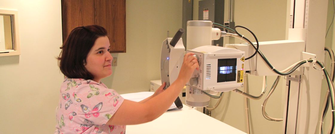
A diagnostic x-ray is a non-invasive medical imaging test used to diagnose a variety of medical conditions, from broken bones to pneumonia. During this quick test, a small dose of radiation is used to create an image of the inside of your body. X-rays can be used to create images of any part of the body.
Our digital X-rays require less radiation to produce images compared to conventional x-ray technology. Your X-ray results will be available quickly to help answer questions about your health. The results are quickly made available to your provider.
X-Rays On Demand
We know that it's difficult to put another appointment in your schedule, that's why we're proud to say that now you don't have to. We welcome walk-ins during our normal business hours. We are inside Bristol Hospital on Level C. Your doctor must order the x-ray before the test can take place. For more information about our services, please call 860.585.3424.
Here at Bristol Hospital we have an accredited Echocardiography Lab, we produce high quality studies with our state of the art machines.
Echocardiography is a valuable imaging test that uses high-frequency sound waves for the diagnosis of structural and functional cardiac disorders. Echocardiograms do not use any radiation.
We do several types of testing:
- Adult Transthoracic Echocardiograms
- 3D and Strain Imaging
- Contrast and Bubble Studies
- Stress Echocardiograms
- Transesophageal Echocardiograms
Advanced Technology with Fewer Risks and Big Benefits
Interventional radiology is one of the fastest growing areas of medicine, combining advanced tools with quality imaging techniques. Many interventional radiology techniques are replacing traditional surgeries because they have lower risks of pain and bleeding and allow you to recover from your procedure much more quickly.
At Bristol Health, our interventional radiologists use high tech imaging and specialized tools like wires and catheters to perform procedures through just one small incision. After your procedure is over, you’ll likely be able to return home the same day and return to all your normal activities quickly.
Because of the low risks of these procedures, most patients are candidates for interventional radiology procedures, despite age or other health conditions. Your doctor will help you learn if an interventional radiology procedure is right for you.
Why It’s Done
Through the use of image-guided technology, our specially trained radiologists are able to perform minimally-invasive procedures to both diagnose and treat, and manage many types of cancer and other conditions.
How the Procedure is Performed
Generally, patients undergoing interventional radiology do not receive general anesthesia and are awake during the procedure. The incision area is made numb with the use of local anesthesia in order to minimize pain and discomfort for the patient.
Interventional radiologists then use imaging techniques such as X-Ray, MRI scans, CT scans and ultrasounds to guide the catheters and instruments to the exact area where the procedure or treatment is to be performed.
The amount of time for recovery varies, but you can rest assured you will feel minimal pain.
Mammograms for Early Detection of Breast Cancer
Mammograms can detect breast cancer in the earliest stages — before it can be felt during a breast exam or cause any noticeable symptoms. It’s important to find cancer early because that’s when it’s most treatable. Early detection and treatment can help prevent the need for more advanced treatment and surgery.
40% Reduced Deaths
Since 1990 according to the American College of Radiology, mammograms have helped reduce deaths from breast cancer in the U.S.
What Is a Mammogram?
A mammogram is a quick, X-ray exam of the breast to look for any abnormalities — this is known as a screening or diagnostic mammogram. Talk to your doctor to find out how often you should have a screening mammogram.
If your doctor notices a change from a previous mammogram or you notice symptoms — such as a lump in your breast, nipple discharge, or unexplained breast pain — they use a diagnostic mammogram to take a closer look.
A mammogram only takes 10 minutes.
3D Mammography
3D mammography, also known as tomosynthesis, is the newest technology in breast imaging. Advanced technology provides physicians with more detailed images of your breast for the most accurate diagnosis.
Studies show that 3D mammography offers improved clarity and cancer detection, especially in women with dense breast tissue. Providing early detection of breast cancer is key to successful treatment and cure, as well as for giving women peace of mind.
Higher Quality Care Means Better Outcomes
The 3D Mammography Unit at Bristol Hospital is FDA-approved and safe for both screening and diagnostic mammograms. Beekley Center is proud to offer this advancement in breast cancer screening as part of our comprehensive breast health and wellness services.
The Beekley Center Difference
At Beekley Center for Breast Health & Wellness, we use the latest in digital breast imaging technology, including 3D mammography (tomosynthesis). We offer the following services:
- A complimentary breast cancer risk assessment during your mammogram. If you’re at greater risk for breast cancer, we offer personalized recommendations to help prevent cancer and monitor your health.
- A welcoming, comfortable environment that’s designed by women, for women.
- Free mammograms for those who are eligible through the Bristol Community Breast Health Project. To find out if you qualify, call us at 860.585.3999.
- Gentle, compassionate care delivered by highly trained mammogram technologists and radiologists.
- Quality you can trust. We’re a Breast Imaging Center of Excellence and accredited by the American College of Radiology, which provides widely accepted breast cancer screening guidelines.
Free mammograms are available for qualified patients. Please contact the Beekley Center at 860.585.3999 for more information and to see if you may qualify.
Advanced Technology. Accurate Diagnosis.
MRI technology uses magnet waves to create detailed images of structures inside your body. These images, particularly of the brain, spine, muscles, and various structures of your anatomy, giving physicians more time and better information to determine the most appropriate treatment.
Convenience and Comfort
At Bristol Health, our MRI unit provides high-quality images with ultra-fast scanning capabilities. Our technologists will work with you to make you comfortable throughout your test. You can also listen to your own music to help keep you calm during your test.
If you experience claustrophobia or are anxious about your MRI, we have anesthesiologists available to provide sedation to help keep you calm and relaxed throughout your test.
How the Test is Performed
Some exams may require a special dye, which, in most cases will be given through a vein (IV) in your hand or forearm. It is used solely to allow the radiologist to see certain things more clearly.
For the MRI, you will lay on a narrow table and be slowly slid into the MRI scanner. During the MRI, the person who operates the machine will watch you from another room. The test lasts about 30 to 60 minutes, but may take longer. You are able to be in contact with the scanner technician during the test.
Our MRI suite is equipped with a stereo for your comfort, please bring your favorite CD or iPod to listen to during your scan.
Wide Bore MRI
We also offer a Wide Bore MRI at the Bristol Radiology Center. This unit is quite larger than a regular MRI providing a comforting feeling for children, older adults, physically larger individuals, or those suffering from claustrophobia. If you are interested in this type of MRI, please call 860.584.0541.
A Safe Way to Study Your Body’s Function
You may be injected with a very small and safe amount of radioactive material to capture images of how your organs work and how they look. This small amount of radiation shows up very clearly in imaging, giving your technologist a highly detailed look at your body. These tests can identify many conditions very early so you can receive more effective treatment.
For your nuclear medicine test, you may be injected with a small amount of radioactive material, and your technologist will then perform imaging of your body. Once your test is over, the material naturally dissipates or is excreted from your body.
Nuclear medicine is particularly unique as it identifies abnormalities very early in the disease process, allowing for immediate intervention and treatment. It is commonly used to diagnose many conditions, including:
- Alzheimer’s disease
- Cancerous tumors throughout the body
- Heart conditions
- Lung conditions
- Thyroid disease
Nuclear Stress Tests
For some, when a routine stress test is unable to determine the cause of their cardiac condition, a nuclear test stress may be needed as well to potentially guide your treatment. A nuclear stress test may be necessary if:
- If you have symptoms such as chest pain or shortness of breath to determine if you have coronary artery disease.
- In helping your doctor determine how well your treatment is working and if any modifications to your treatment plan need to be made.
Highly Detailed and Accurate Imaging
Positron emission tomography (PET) is a nuclear medicine test that creates pictures of how your body works. It is most often used for cancer diagnosis and monitoring, though it can also be used to assess brain and heart conditions.
By reflecting small changes in your cells, PET scans detect disease early on and help you and your physician know if cancer has spread. PET scans can tell the location of a tumor, whether the tumor is benign or malignant, whether the cancer has spread, and whether treatment has been effective.
Combined With CT Scan
A computed tomography (CT) scan produces detailed images that reveal the size and location of a tumor. Combining these two scanning technologies helps your physician see a more detailed and accurate picture of your health.
PET/CT Scans and Cancers
The PET/CT scanner is able to “see” damaged or cancerous cells where the injected glucose substance is being absorbed. Cancer cells use more glucose than do normal cells. The rate in which a tumor is using the glucose helps determine the level and aggressiveness of the cancer.
The PET/CT scan provides a more detailed picture of cancerous tissues than either test does on its own. The images are captured in a single scan.
The Safest Imaging Technology
Ultrasound doesn’t use any radiation. The technology uses high-frequency sound waves to view live images of the organs, tissues, and blood vessels inside your body.
Ultrasound is so safe that it is routinely used during pregnancy. It is also an extremely useful way to examine internal organs such as the liver, gallbladder, spleen, pancreas, kidney, scrotum, pelvis, breast, thyroid, and vascular system.
Maternal-Fetal Ultrasound
During a maternal health ultrasound, a perinatologist (a doctor who specializes in maternal-fetal medicine) very carefully examines the fetus using advanced ultrasound technology. These ultrasounds allow your perinatologist to diagnose conditions in your child before birth.
Obstetrics Ultrasound
Throughout your pregnancy, you will receive ultrasounds to check on your baby’s growth as well as the health of your placenta, uterus, and cervix.
Vascular Ultrasound
Vascular ultrasounds allow your doctors to study how your blood flows and identify any areas where blood flow is decreased.
Breast Ultrasound
Along with other testing, breast ultrasound can be used to help determine if lumps in the breast are benign or cancerous.
